hero image

Digital PCR assays for melanoma gene variants

Order ready-to-use dPCR assays

Revolutionizing melanoma research with precision dPCR assays

Melanoma research is filled with challenges, due to the complexity and diversity of tumor heterogeneity, which complicates the development of effective treatments. Analysis of challenging samples, such as those with low tumor purity or from metastatic sites, further hinders progress.

Digital PCR is emerging as a valuable technology that can help address these issues by providing highly sensitive and precise detection of genetic variations. This technology enhances researchers' ability to accurately profile tumors, even from minimal or poor-quality samples. Consequently, digital PCR holds promise for advancing melanoma research by enabling better understanding and targeting of the disease at its earliest and most treatable stages.

Explore melanoma related dPCR assays by gene

Melanoma’s genetic complexity plays a fundamental role in both its initiation and progression. The disease arises through multiple genetic pathways, with key mutations that profoundly impact both the clinical trajectory and therapeutic approaches. Central to melanoma oncogenesis are mutations in the BRAF, NRAS and CDKN2A genes. BRAF mutations, especially the V600E variant, are particularly significant in early melanoma stages, driving abnormal cell growth and proliferation through the MAPK/ERK pathway. NRAS mutations influence cell signaling and are associated with more aggressive disease and resistance to some therapies. CDKN2A mutations disrupt cell cycle regulation, contributing to uncontrolled cell division and tumor progression.

Our collection of dPCR LNA Mutation Assays offer a comprehensive toolkit for researchers dedicated to dissecting these critical genetic alterations. By enabling the precise detection and quantification of these key mutations, our assays support the development of targeted research and therapies, paving the way for more personalized and effective treatment approaches.

The table below includes COSMIC Variant IDs (COSV), as they provide a unique identifier for each distinct gene mutation and ensure precise reference to your variant of interest.
Gene
Mutation Type
Gene
Mutation Type
Mutation (CDS)
Mutation (AA)
COSMIC ID (COSV)
COSMIC ID (COSM)
Codon
dPCR Mutation Assay
BRAF Substitution - Missensec.1406G>Cp.G469ACOSV56061424COSM460469 DMH0000047
BRAF Substitution - Missensec.1798G>Ap.V600MCOSV56075762COSM1130600 DMH0000218
BRAF Substitution - Missensec.1798_1799delinsAAp.V600KCOSV56057713COSM473600 DMH0000001
BRAF Substitution - Missensec.1798_1799delinsAGp.V600RCOSV56058419COSM474600 DMH0000002
BRAF Substitution - Missensec.1799T>Ap.V600ECOSV56056643COSM476600 DMH0000004
BRAF Substitution - Missensec.1799T>Gp.V600GCOSV56080151COSM6137600 DMH0000068
BRAF Substitution - Missensec.1799_1800delinsAAp.V600ECOSV56059110COSM475600 DMH0000003
BRAF Substitution - Missensec.1799_1800delinsATp.V600DCOSV56059623COSM477600 DMH0000039
EGFR Substitution - Missensec.2573T>Gp.L858RCOSV51765161COSM6224858 DMH0000386
IDH1 Substitution - Missensec.394C>Ap.R132SCOSV61615649COSM28748132 DMH0000066
IDH1 Substitution - Missensec.394C>Gp.R132GCOSV61615456COSM28749132 DMH0000063
IDH1 Substitution - Missensec.394_395delinsGTp.R132VCOSV61616571COSM28751132 DMH0000228
IDH1 Substitution - Missensec.395G>Tp.R132LCOSV61615420COSM28750132 DMH0000015
KRAS Substitution - Missensec.34G>Ap.G12SCOSV55497461COSM51712 DMH0000519
KRAS Substitution - Missensec.34G>Cp.G12RCOSV55497582COSM51812 DMH0000284
KRAS Substitution - Missensec.34G>Tp.G12CCOSV55497469COSM51612 DMH0000309
KRAS Substitution - Missensec.35G>Ap.G12DCOSV55497369COSM52112 DMH0000286
KRAS Substitution - Missensec.35G>Cp.G12ACOSV55497479COSM52212 DMH0001055
KRAS Substitution - Missensec.35G>Tp.G12VCOSV55497419COSM52012 DMH0000285
KRAS Substitution - Missensec.37G>Ap.G13SCOSV55509530COSM52813 DMH0000331
KRAS Substitution - Missensec.37G>Cp.G13RCOSV55502117COSM52913 DMH0000332
KRAS Substitution - Missensec.37G>Tp.G13CCOSV55497378COSM52713 DMH0000195
KRAS Substitution - Missensec.38G>Ap.G13DCOSV55497388COSM53213 DMH0000289
KRAS Substitution - Missensec.38G>Cp.G13ACOSV55497357COSM53313 DMH0000334
KRAS Substitution - Missensec.38G>Tp.G13VCOSV55522580COSM53413 DMH0000527
KRAS Substitution - Missensec.38_39delinsATp.G13DCOSV55508630COSM53113 DMH0000525
NRAS Substitution - Missensec.34G>Ap.G12SCOSV54736621COSM56312 DMH0000188
NRAS Substitution - Missensec.34G>Cp.G12RCOSV54736940COSM56112 DMH0000336
NRAS Substitution - Missensec.34G>Tp.G12CCOSV54736487COSM56212 DMH0000186
NRAS Substitution - Missensec.35G>Cp.G12ACOSV54736555COSM56512 DMH0000339
NRAS Substitution - Missensec.35G>Tp.G12VCOSV54736974COSM56612 DMH0000340
NRAS Substitution - Missensec.37G>Ap.G13SCOSV54736476COSM57113 DMH0000510
NRAS Substitution - Missensec.37G>Cp.G13RCOSV54736550COSM56913 DMH0000341
NRAS Substitution - Missensec.37G>Tp.G13CCOSV54736386COSM57013 DMH0000342
NRAS Substitution - Missensec.38G>Ap.G13DCOSV54736416COSM57313 DMH0000343
NRAS Substitution - Missensec.38G>Cp.G13ACOSV54736793COSM57513 DMH0000345
NRAS Substitution - Missensec.38G>Tp.G13VCOSV54736480COSM57413 DMH0000344
NRAS Substitution - Missensec.181C>Ap.Q61KCOSV54736310COSM58061 DMH0000505
NRAS Substitution - Missensec.181C>Gp.Q61ECOSV54743343COSM58161 DMH0000347
NRAS Substitution - Missensec.182A>Gp.Q61RCOSV54736340COSM58461 DMH0000183
NRAS Substitution - Missensec.182A>Tp.Q61LCOSV54736624COSM58361 DMH0000190
NRAS Substitution - Missensec.183A>Cp.Q61HCOSV54736320COSM58661 DMH0000180
NRAS Substitution - Missensec.183A>Tp.Q61HCOSV54736991COSM58561 DMH0000349
PIK3CA Substitution - Missensec.1633G>Ap.E545KCOSV55873239COSM763545 DMH0000292
PIK3CA Substitution - Missensec.1634A>Gp.E545GCOSV55873220COSM764545 DMH0000033
PIK3CA Substitution - Missensec.1636C>Ap.Q546KCOSV55873527COSM766546 DMH0000037
PIK3CA Substitution - Missensec.1637A>Gp.Q546RCOSV55876869COSM12459546 DMH0000212
PIK3CA Substitution - Missensec.3139C>Tp.H1047YCOSV55876499COSM7741047 DMH0000209
PIK3CA Substitution - Missensec.3140A>Gp.H1047RCOSV55873195COSM7751047 DMH0000036
PIK3CA Substitution - Missensec.3140A>Tp.H1047LCOSV55873401COSM7761047 DMH0000062
PTEN Substitution - Missensec.388C>Gp.R130GCOSV64288384COSM5219130 DMH0000296
PTEN Substitution - Nonsensec.388C>Tp.R130*COSV64288463COSM5152130 DMH0000294
TP53 Substitution - Missensec.488A>Gp.Y163CCOSV52663142COSM10808163 DMH0000112
TP53 Substitution - Missensec.517G>Tp.V173LCOSV52676535COSM43559173 DMH0000126
TP53 Substitution - Missensec.659A>Gp.Y220CCOSV52661282COSM10758220 DMH0000440
TP53 Substitution - Missensec.743G>Tp.R248LCOSV52675468COSM6549248 DMH0000381
TP53 Substitution - Missensec.818G>Ap.R273HCOSV52660980COSM10660273 DMH0000094
TP53 Substitution - Missensec.818G>Tp.R273LCOSV52664805COSM10779273 DMH0000114
TP53 Substitution - Missensec.833C>Tp.P278LCOSV52678063COSM10863278 DMH0000372
TP53 Substitution - Missensec.856G>Ap.E286KCOSV52664318COSM10726286 DMH0000364
Items per page:
1 – 60 of 60

Discover the QIAcuity family of dPCR instruments

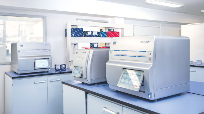
Explore the QIAcuity family of dPCR instruments, designed to meet the rigorous demands of biomarker research, translational research and clinical applications. With unmatched precision and operational efficiency, these versatile platforms can transform your scientific and diagnostic efforts.

Transform your research capabilities with QIAcuity digital PCR

QIAcuity is a fully automated digital PCR system that combines precision, efficiency and ease of use. Experience unparalleled accuracy and save resources with high-throughput multiplexing, allowing for the simultaneous detection of up to five genetic targets. A seamless transition from existing qPCR workflows ensures minimal disruption while significantly enhancing data quality and throughput.

Streamline your clinical PCR workflows with QIAcuityDx

QIAcuityDx is tailored for IVD applications. This fully automated system enhances diagnostic precision and operational efficiency by reducing hands-on time and ensuring accurate detection and quantification of important genetic variations. Easily develop your own assay menu* by using QIAcuityDx utility mode and IVD medical device consumables, reagents and software.

*FDA ‘Medical Devices; Laboratory Developed Tests’ final rule, May 6, 2024 and European Union regulation requirements on ‘In-House Assays’ (Regulation (EU) 2017/746 -IVDR- Art. 5(5))

Frequently asked questions

Discover key insights into genes and variants critical for melanoma research and how they can be detected using digital PCR

How do dPCR LNA Mutation Assays benefit cancer researchers?

dPCR LNA Mutation Assays offer significant advantages to cancer researchers working on precise and sensitive mutation detection. These assays are specifically designed for use with the QIAcuity Digital PCR System and are enhanced with Locked Nucleic Acid (LNA) technology. This enhancement greatly improves the specificity and sensitivity of mutation detection, making it possible to identify DNA sequence mutations at very low abundance, with a sensitivity as fine as 0.1% in a single nanoplate well.

The key benefits of dPCR LNA Mutation Assays for cancer researchers include:
  • High precision and sensitivity: The use of duplex, hydrolysis probe-based assays allows for highly precise detection of mutations. The presence of both mutant and wild-type probes in the same reaction ensures that researchers can detect and quantify minor genetic variations with great accuracy, crucial for studies in heterogeneous cancer samples where only a few cells may carry the mutation.
  • Enhanced specificity: The integration of LNA into the probes increases the binding affinity and specificity towards the target sequences, minimizing the risk of non-specific bindings and improving the overall reliability of the assays.
  • Multiplexing capability: Each assay is capable of detecting mutations using two different fluorescent dye combinations, allowing for the simultaneous analysis of mutant and wild-type alleles within the same reaction. This multiplexing ability is particularly useful in applications requiring the analysis of multiple targets, such as assessing co-occurring mutations in cancer.
  • Flexibility in sample analysis: By dividing the reaction across multiple wells, even greater sensitivity can be achieved, facilitating the detection of extremely rare mutations. This is especially valuable in cancer research, where detecting low-frequency mutations can inform prognosis and treatment strategies.
  • Streamlined workflow: Supplied in a single-tube format with ready-to-use primer pairs and probes, these assays simplify the experimental setup, enabling efficient and straightforward integration into existing research workflows.

What role does BRAF play in melanoma?

BRAF (v-raf murine sarcoma viral oncogene homolog B) is a gene that encodes a protein known as B-Raf, which is a serine/threonine-protein kinase. This protein is part of the MAPK signaling pathway, which plays a crucial role in regulating cell division, differentiation and secretion. BRAF interacts with other proteins in the MAPK pathway, including MEK and ERK, to transmit signals from the cell surface to the DNA in the cell nucleus. In melanoma, BRAF mutations lead to the continuous activation of the MAPK pathway, promoting uncontrolled cell growth and survival. This makes BRAF a significant target for therapeutic interventions in melanoma. Common BRAF-associated variants include the following:
  • BRAF c.1799T>A / V600E: A DNA point mutation from thymine (T) to adenine (A) at nucleotide position 1799 (c.1799T>A) results in the substitution of valine (V) with glutamic acid (E) at position 600 of the BRAF protein (V600E). This mutation leads to a constitutively active BRAF kinase, which continuously signals through the MAPK/ERK pathway without the need for external growth signals. This results in increased cell proliferation and survival, contributing to melanoma development and progression. The V600E mutation is the most common BRAF mutation in melanoma, found in approximately 50-60% of cases. It is a key target for BRAF inhibitors like vemurafenib and dabrafenib, which have shown significant efficacy in treating BRAF V600E-mutant melanoma.
  • BRAF c.1798_1799GT>AA / V600K: A DNA mutation involving the substitution of guanine (G) and thymine (T) with adenine (A) and adenine (A) at nucleotide positions 1798 and 1799 (c.1798_1799GT>AA) results in the substitution of valine (V) with lysine (K) at position 600 of the BRAF protein (V600K). Similar to V600E, this mutation leads to constitutive activation of the BRAF kinase and the MAPK/ERK pathway, promoting melanoma cell growth and survival. The V600K mutation is the second most common BRAF mutation in melanoma, found in about 10-30% of cases. Patients with this mutation also benefit from BRAF inhibitors, although the response rates and progression-free survival may differ slightly from those with V600E mutations.
  • BRAF c.1798_1799GT>AG / V600R: A DNA mutation from guanine (G) and thymine (T) to adenine (A) and guanine (G) at nucleotide positions 1798 and 1799 (c.1798_1799GT>AG) results in the substitution of valine (V) with arginine (R) at position 600 of the BRAF protein (V600R). This mutation results in constitutive activation of the BRAF kinase, leading to continuous MAPK/ERK pathway signaling and promoting melanoma cell proliferation and survival. The V600R mutation is rare, accounting for about 0.9-5% of BRAF-mutant melanomas. It responds to BRAF inhibitors, although the clinical outcomes and response rates may vary compared to V600E and V600K mutations.
  • BRAF Splicing Variants: BRAF splicing variants, such as p61, p55, p48 and p41, result from alternative splicing events that produce truncated forms of the BRAF protein. These splicing variants can lead to resistance to BRAF inhibitors by maintaining MAPK/ERK pathway activation even in the presence of these drugs. They often emerge during treatment and contribute to disease progression. Detection of these splicing variants in circulating tumor DNA (ctDNA) or extracellular vesicles (EVs) can provide insights into resistance mechanisms and guide adjustments in therapeutic strategies.

What is the significance of CDKN2A in melanoma?

CDKN2A (cyclin-dependent kinase inhibitor 2A) is a tumor suppressor gene that encodes two proteins, p16INK4a and p14ARF, through alternative splicing. These proteins play a role in regulating the cell cycle. p16INK4a inhibits cyclin-dependent kinases 4 and 6 (CDK4/6), preventing the phosphorylation of the retinoblastoma protein (Rb) and halting cell cycle progression from G1 to S phase. p14ARF stabilizes the tumor suppressor protein p53 by inhibiting its degradation. In melanoma, loss of CDKN2A function leads to uncontrolled cell division and contributes to tumorigenesis. CDKN2A is a critical regulator of cell cycle checkpoints and apoptosis. Some CDKN2A variants relevant to melanoma include:
  • CDKN2A c.256G>A / G86R: A DNA point mutation from guanine (G) to adenine (A) at nucleotide position 256 (c.256G>A) results in the substitution of glycine (G) with arginine (R) at position 86 of the CDKN2A protein (G86R). This mutation disrupts the function of the p16INK4a protein, which normally inhibits cyclin-dependent kinases 4 and 6 (CDK4/6), leading to uncontrolled cell cycle progression. The loss of p16INK4a function results in increased cell proliferation and survival, contributing to melanoma development and progression. The G86R mutation is one of the pathogenic variants in CDKN2A associated with a high risk of melanoma, particularly in familial cases.
  • CDKN2A c.301G>T / G101W: A DNA point mutation from guanine (G) to thymine (T) at nucleotide position 301 (c.301G>T) results in the substitution of glycine (G) with tryptophan (W) at position 101 of the CDKN2A protein (G101W). This mutation impairs the ability of p16INK4a to bind and inhibit CDK4/6, leading to deregulated cell cycle progression and increased melanoma risk. The G101W mutation is another significant variant in CDKN2A, often found in patients with a family history of melanoma and other cancers, such as pancreatic cancer.
  • CDKN2A c.377_378del / p.Pro126fs: A deletion of two nucleotides, cytosine (C) and thymine (T), at positions 377 and 378 (c.377_378del) results in a frameshift mutation, leading to a premature stop codon and truncation of the CDKN2A protein (p.Pro126fs). This truncation eliminates critical functional domains of p16INK4a, rendering it unable to inhibit CDK4/6. The loss of functional p16INK4a promotes uncontrolled cell division and contributes to melanoma development. The p.Pro126fs mutation is associated with a high risk of melanoma and is often identified in familial melanoma cases.
  • CDKN2A c.442G>A / R148H: A DNA point mutation from guanine (G) to adenine (A) at nucleotide position 442 (c.442G>A) results in the substitution of arginine (R) with histidine (H) at position 148 of the CDKN2A protein (R148H). This mutation affects the p14ARF isoform of CDKN2A, which plays a role in stabilizing the tumor suppressor protein p53. The R148H mutation disrupts p14ARF function, leading to decreased p53 activity and impaired cell cycle arrest and apoptosis. This contributes to increased melanoma risk and progression. The R148H mutation is less common but still significant in the context of melanoma susceptibility.

How is GNA11 involved in melanoma?

GNA11 (G protein subunit alpha 11) is a gene that encodes a G protein alpha subunit similar to GNAQ, involved in the activation of PLC and downstream signaling pathways. GNA11 plays a role in cell proliferation, differentiation and survival through the activation of PKC and the MAPK pathway. In uveal melanoma, mutations in GNA11 lead to constitutive activation of these pathways, driving tumor growth and progression.

What is the role of GNAQ in melanoma?

GNAQ (G protein subunit alpha q) is a gene that encodes a G protein alpha subunit involved in the activation of phospholipase C (PLC) and subsequent intracellular signaling pathways. GNAQ plays a role in regulating cell proliferation, differentiation and survival through the activation of downstream effectors such as protein kinase C (PKC) and the MAPK pathway. In melanoma, particularly uveal melanoma, mutations in GNAQ lead to constitutive activation of these signaling pathways, promoting tumor growth and survival.

How does KIT affect melanoma?

KIT (KIT proto-oncogene receptor tyrosine kinase) is a gene that encodes a receptor tyrosine kinase known as c-Kit or CD117. This receptor is involved in various cellular processes, including cell survival, proliferation and differentiation. KIT interacts with its ligand, stem cell factor (SCF), to activate downstream signaling pathways such as MAPK, PI3K/AKT and JAK/STAT. In melanoma, particularly in mucosal and acral subtypes, KIT mutations or amplifications can lead to constitutive activation of these pathways, driving tumor growth and survival. KIT is a target for specific inhibitors in melanoma therapy.

What role does MITF play in melanoma?

MITF (microphthalmia-associated transcription factor) is a transcription factor that plays a key role in melanocyte development, function and survival. It regulates the expression of genes involved in melanin production, cell cycle progression and apoptosis. MITF interacts with various signaling pathways, including MAPK and PI3K/AKT, to control melanocyte differentiation and proliferation. In melanoma, dysregulation of MITF can contribute to tumorigenesis by promoting cell survival and resistance to apoptosis. MITF is considered a lineage-specific oncogene in melanoma.

How is NRAS involved in melanoma?

NRAS (neuroblastoma RAS viral oncogene homolog) is a gene that encodes a protein called N-Ras, which is a small GTPase involved in the MAPK and PI3K/AKT signaling pathways. These pathways are essential for cell proliferation, differentiation and survival. NRAS interacts with various downstream effectors, including RAF kinases, PI3K and RalGDS, to propagate signals that control cell growth and apoptosis. In melanoma, NRAS mutations can lead to the activation of these signaling pathways, contributing to tumor development and progression. NRAS is also implicated in resistance to certain therapies, making it a critical focus in melanoma research. Some significant NRAS variants include:
  • NRAS c.182A>G / Q61R: A DNA point mutation from adenine (A) to guanine (G) at nucleotide position 182 (c.182A>G) results in the substitution of glutamine (Q) with arginine (R) at position 61 of the NRAS protein (Q61R). This mutation results in a constitutively active NRAS protein, which continuously signals through the MAPK/ERK and PI3K/AKT pathways without the need for external growth signals. This leads to increased cell proliferation, survival and migration, contributing to melanoma development and progression. The Q61R mutation is the most common NRAS mutation in melanoma, found in approximately 80-90% of NRAS-mutant cases. It is associated with a more aggressive disease phenotype and poorer prognosis compared to other NRAS mutations.
  • NRAS c.181C>A / Q61K: A DNA point mutation from cytosine (C) to adenine (A) at nucleotide position 181 (c.181C>A) results in the substitution of glutamine (Q) with lysine (K) at position 61 of the NRAS protein (Q61K). Similar to Q61R, this mutation leads to constitutive activation of NRAS, driving continuous MAPK/ERK and PI3K/AKT pathway signaling. This promotes melanoma cell growth, survival and metastasis. The Q61K mutation is less common than Q61R but still significant, contributing to the aggressive behavior of NRAS-mutant melanomas. It is found in a smaller subset of NRAS-mutant melanomas and is associated with resistance to certain therapies.
  • NRAS c.182A>T / Q61L: A DNA point mutation from adenine (A) to thymine (T) at nucleotide position 182 (c.182A>T) results in the substitution of glutamine (Q) with leucine (L) at position 61 of the NRAS protein (Q61L). This mutation also results in constitutive activation of NRAS, leading to persistent MAPK/ERK and PI3K/AKT pathway signaling. This enhances melanoma cell proliferation, survival and invasiveness. The Q61L mutation is relatively rare compared to Q61R and Q61K but still plays a role in melanoma pathogenesis. It is associated with a distinct biological behavior and may influence the response to targeted therapies.
  • NRAS c.35G>A / G12D: A DNA point mutation from guanine (G) to adenine (A) at nucleotide position 35 (c.35G>A) results in the substitution of glycine (G) with aspartic acid (D) at position 12 of the NRAS protein (G12D). This mutation leads to a constitutively active NRAS protein, which continuously activates downstream signaling pathways, including MAPK/ERK and PI3K/AKT. This promotes melanoma cell growth and survival. The G12D mutation is less common in melanoma compared to Q61 mutations but still contributes to the oncogenic potential of NRAS. It is associated with a different set of clinical and pathological features compared to Q61 mutations.

How do PTEN mutations contribute to melanoma?

PTEN (phosphatase and tensin homolog) is a tumor suppressor gene that encodes a protein with lipid phosphatase activity, which negatively regulates the PI3K/AKT signaling pathway. PTEN dephosphorylates phosphatidylinositol (3,4,5)-trisphosphate (PIP3), thereby inhibiting AKT activation and promoting apoptosis and cell cycle arrest. In melanoma, loss or downregulation of PTEN leads to increased PI3K/AKT pathway activity, promoting cell survival, proliferation and migration. PTEN also interacts with other signaling pathways, including MAPK, and its loss is associated with poor prognosis in melanoma.

How do TERT mutations contribute to melanoma?

TERT (telomerase reverse transcriptase) is a gene that encodes the catalytic subunit of telomerase, an enzyme responsible for maintaining telomere length. Telomeres protect chromosome ends from degradation and fusion, and their maintenance is crucial for cellular immortality. In melanoma, upregulation of TERT activity allows for continuous cell division and tumor growth. TERT interacts with various proteins involved in telomere maintenance and DNA repair. Mutations in the TERT promoter can lead to increased expression and are associated with poor prognosis in melanoma.

What impact does TP53 have on melanoma?

TP53 (tumor protein p53) is a tumor suppressor gene that encodes the p53 protein, which is crucial for maintaining genomic stability. p53 functions as a transcription factor that regulates the expression of genes involved in cell cycle arrest, DNA repair, apoptosis and senescence. In response to cellular stress or DNA damage, p53 activates pathways that prevent the propagation of damaged cells. In melanoma, TP53 mutations can lead to the loss of these protective mechanisms, allowing for uncontrolled cell growth and survival. p53 interacts with various proteins, including MDM2, which regulates its stability and activity.

Disclaimers

dPCR LNA Mutation Assays are intended for molecular biology applications. These products are not intended for the diagnosis, prevention, or treatment of a disease.

The QIAcuity is intended for molecular biology applications. This product is not intended for the diagnosis, prevention or treatment of a disease. Therefore, the performance characteristics of the product for clinical use (i.e., diagnostic, prognostic, therapeutic or blood banking) is unknown.

The QIAcuityDx dPCR System is intended for in vitro diagnostic use, using automated multiplex quantification dPCR technology, for the purpose of providing diagnostic information concerning pathological states.

QIAcuity and QIAcuityDx dPCR instruments are sold under license from Bio-Rad Laboratories, Inc. and exclude rights for use with pediatric applications. The QIAcuityDx medical device is currently under development and will be available in 20 countries in H2 2024.