hero image

Digital PCR assays for gene variants related to acute leukemias

Order ready-to-use dPCR assays

Revolutionizing acute leukemia research with precision dPCR assays

Acute leukemia research faces significant challenges due to the genetic diversity and complexity of these diseases, with numerous subtypes and mutations making it difficult to develop universal treatments and identify reliable biomarkers. Plus, clonal evolution allows for new mutations over time, particularly under treatment pressure, leading to drug resistance. Minimal residual disease (MRD) detection is also a challenge, as detecting the small number of leukemic cells requires highly sensitive techniques.

Digital PCR (dPCR) offers promising solutions with its ultra-sensitive detection capabilities, enabling researchers to identify and quantify rare genetic mutations and variants associated with acute leukemias. This precision allows for the detection of low-frequency mutations, tracking clonal evolution and MRD, providing insights into the genetic diversity and complexity of the disease.

Explore dPCR assays related to acute leukemias

Acute leukemias, such as acute myeloid leukemia (AML) and acute lymphoblastic leukemia (ALL), are diverse diseases, each marked by distinct genetic characteristics and treatment responses. Understanding key mutations in genes like FLT3, NPM1, DNMT3A, IDH1 and IDH2 is fundamental in advancing our knowledge of disease progression and identifying potential therapeutic targets. These insights are also critical for studying treatment resistance mechanisms and developing more effective targeted therapies.

Our collection of dPCR LNA Mutation Assays provides a robust toolkit for advanced hematology research, facilitating precise genetic analysis and insights.

The table below includes COSMIC Variant IDs (COSV), as they provide a unique identifier for each distinct gene mutation and ensure precise reference to your variant of interest.

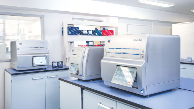
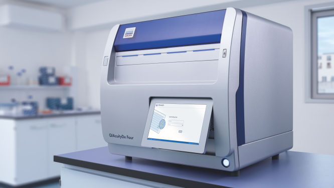
Discover the QIAcuity family of dPCR instruments

Explore the QIAcuity family of dPCR instruments, designed to meet the rigorous demands of biomarker research, translational research and clinical applications. With unmatched precision and operational efficiency, these versatile platforms can transform your scientific and diagnostic efforts.

Transform your research capabilities with QIAcuity digital PCR

QIAcuity is a fully automated digital PCR system that combines precision, efficiency and ease of use. Experience unparalleled accuracy and save resources with high-throughput multiplexing, allowing for the simultaneous detection of up to five genetic targets. A seamless transition from existing qPCR workflows ensures minimal disruption while significantly enhancing data quality and throughput.

Streamline your clinical PCR workflows with QIAcuityDx

QIAcuityDx is tailored for IVD applications. This fully automated system enhances diagnostic precision and operational efficiency by reducing hands-on time and ensuring accurate detection and quantification of important genetic variations. Easily develop your own assay menu* by using QIAcuityDx utility mode and IVD medical device consumables, reagents and software.

*FDA ‘Medical Devices; Laboratory Developed Tests’ final rule, May 6, 2024 and European Union regulation requirements on ‘In-House Assays’ (Regulation (EU) 2017/746 -IVDR- Art. 5(5))

Frequently asked questions

Discover key insights into genes and variants critical for acute leukemia research and how they can be detected using digital PCR
How do dPCR LNA Mutation Assays benefit cancer researchers?
dPCR LNA Mutation Assays offer significant advantages to cancer researchers requiring precise and sensitive mutation detection. These assays are specifically designed for use with the QIAcuity Digital PCR System and are enhanced with Locked Nucleic Acid (LNA) technology. This enhancement greatly improves the specificity and sensitivity of mutation detection, making it possible to identify DNA sequence mutations at very low abundance, with a sensitivity as fine as 0.1% in a single nanoplate well.

The key benefits of dPCR LNA Mutation Assays for cancer researchers include:
  • High precision and sensitivity: The use of duplex, hydrolysis probe-based assays allows for highly precise detection of mutations. The presence of both mutant and wild-type probes in the same reaction ensures that researchers can detect and quantify minor genetic variations with great accuracy, crucial for studies in heterogeneous cancer samples where only a few cells may carry the mutation.
  • Enhanced specificity: The integration of LNA into the probes increases the binding affinity and specificity towards the target sequences, minimizing the risk of non-specific bindings and improving the overall reliability of the assays.
  • Multiplexing capability: Each assay is capable of detecting mutations using two different fluorescent dye combinations, allowing for the simultaneous analysis of mutant and wild-type alleles within the same reaction. This multiplexing ability is particularly useful in applications requiring the analysis of multiple targets, such as assessing co-occurring mutations in cancer.
  • Flexibility in sample analysis: By dividing the reaction across multiple wells, even greater sensitivity can be achieved, facilitating the detection of extremely rare mutations. This is especially valuable in cancer research, where detecting low-frequency mutations can inform prognosis and treatment strategies.
  • Streamlined workflow: Supplied in a single-tube format with ready-to-use primer pairs and probes, these assays simplify the experimental setup, enabling efficient and straightforward integration into existing research workflows.
What role does the CEBPA gene play in acute myeloid leukemia?
The CEBPA gene encodes the CCAAT/enhancer-binding protein alpha, a transcription factor that is crucial for granulocyte differentiation and function. CEBPA regulates the expression of genes involved in the proliferation and differentiation of myeloid progenitor cells into granulocytes. It functions by binding to specific DNA sequences in the promoter regions of target genes, thereby activating their transcription. CEBPA also interacts with other transcription factors and co-factors to form a regulatory network that ensures the proper development of granulocytes. Mutations in CEBPA, particularly double mutations, impair its DNA binding and transcriptional activation capabilities, leading to a block in granulocyte differentiation and the accumulation of immature myeloid cells. In acute myeloid leukemia (AML), patients with CEBPA double mutations often have a better prognosis compared to other AML subtypes.
What is the significance of the DNMT3A gene in acute leukemias?
DNMT3A (DNA Methyltransferase 3 Alpha) is an enzyme responsible for de novo DNA methylation, playing a key role in gene regulation and maintaining genomic stability. DNMT3A mutations are commonly observed in AML, with about 20–30% of AML patients harboring these mutations. They are also found in a smaller percentage of ALL cases. The R882H mutation is a hotspot that significantly impacts the enzyme's function, leading to altered methylation patterns and contributing to leukemogenesis. DNMT3A mutations often co-occur with other genetic alterations, compounding their impact on disease progression. Understanding these mutations is crucial for developing therapeutic strategies aimed at correcting aberrant methylation.
  • DNMT3A c.2645G>A / p.R882H: A hotspot mutation at nucleotide 2645 from guanine (G) to adenine (A) results in the substitution of arginine (R) with histidine (H) at position 882. This mutation disrupts the enzyme's methyltransferase activity due to alteration in the catalytic domain, leading to widespread hypomethylation and the activation of oncogenes such as FLT3 and NRAS. The R882H mutation also promotes clonal expansion of hematopoietic stem cells with leukemic potential. It is associated with poor prognosis and resistance to standard chemotherapy, necessitating the development of new therapeutic approaches targeting these epigenetic changes.
  • DNMT3A c.2644G>T / p.R882C: A point mutation at nucleotide 2644 from guanine (G) to thymine (T) results in the substitution of arginine (R) with cysteine (C) at position 882. This mutation disrupts DNMT3A's methyltransferase activity, leading to epigenetic alterations such as hypomethylation and aberrant activation of oncogenes, including MYC and HOXA9, which drive leukemogenesis, where normal hematopoietic cells transform into leukemic cells. The R882C mutation impairs the enzyme's ability to maintain normal methylation patterns, contributing to the malignant transformation of hematopoietic cells. This mutation is also associated with poor prognosis, highlighting the need for therapeutic strategies that address these specific epigenetic changes.
  • DNMT3A c.2104A>G / p.Y735C: A point mutation at nucleotide 2104 from adenine (A) to guanine (G) results in the substitution of tyrosine (Y) with cysteine (C) at position 735. This mutation occurs in the catalytic domain of DNMT3A, impairing its ability to bind to DNA and execute its methyltransferase function. The Y735C mutation leads to abnormal methylation patterns of genes such as CDKN2A and P15, and the deregulation of gene expression, including genes like AML1 and CEBPA, promoting the development of leukemia. The presence of this mutation can contribute to treatment resistance, underscoring the importance of epigenetic therapy in acute myeloid leukemia.
  • DNMT3A c.2150G>A / p.R750H: A point mutation at nucleotide 2150 from guanine (G) to adenine (A) results in the substitution of arginine (R) with histidine (H) at position 750. This mutation affects the enzyme’s structural integrity and its interaction with DNA, leading to reduced methyltransferase activity. The R750H mutation causes widespread epigenetic dysregulation, contributing to the clonal expansion of leukemic cells. It is associated with poor prognosis and necessitates the development of novel therapeutic approaches that can mitigate its impact on gene expression and cell proliferation.
What role does the FLT3 gene play in acute myeloid leukemia?
The FLT3 gene encodes a receptor tyrosine kinase that is crucial for the normal development of hematopoietic stem cells. Mutations in FLT3, particularly internal tandem duplications (ITD) and point mutations in the tyrosine kinase domain (TKD), are among the most common in acute myeloid leukemia (AML) and are associated with poor prognosis. FLT3-ITD mutations result in constitutive activation of the FLT3 receptor, leading to uncontrolled cell proliferation and survival. The continuous activation of signaling pathways such as the PI3K/AKT and RAS/MAPK pathways promotes leukemogenesis. Understanding FLT3 mutations is essential for developing targeted therapies, such as FLT3 inhibitors, which have shown promise in improving outcomes for AML patients.
  • FLT3-ITD (Internal Tandem Duplication): This mutation involves the insertion of duplicated sequences in the juxtamembrane domain of the FLT3 receptor. This duplication leads to constitutive activation of the receptor, independent of its ligand, resulting in persistent signaling through pathways such as PI3K/AKT and RAS/MAPK. This unchecked signaling promotes proliferation and survival of leukemic cells, contributing to the aggressive nature of AML. The FLT3-ITD mutation is also associated with an increased risk of relapse and resistance to conventional chemotherapy, making it a critical target for new therapeutic approaches.
  • FLT3 c.2503G>T / p.D835Y: A point mutation at nucleotide 2503 from guanine (G) to thymine (T) results in the substitution of aspartic acid (D) with tyrosine (Y) at position 835 in the tyrosine kinase domain (TKD) of FLT3. This mutation alters the kinase's conformation, leading to constitutive activation of the receptor. The D835Y mutation disrupts normal kinase regulation, enhancing downstream signaling pathways such as PI3K/AKT, RAS/MAPK and STAT5, which promote cell proliferation and survival. This mutation is often linked to resistance to FLT3 inhibitors, necessitating the development of second-generation inhibitors that can effectively target this variant.
  • FLT3 c.1804C>T / p.A602V: A point mutation at nucleotide 1804 from cytosine (C) to thymine (T) results in the substitution of alanine (A) with valine (V) at position 602 in the juxtamembrane domain of FLT3. This mutation affects the stability of the FLT3 receptor and can lead to its constitutive activation. The A602V mutation enhances downstream signaling pathways such as PI3K/AKT and RAS/MAPK, promoting the proliferation and survival of leukemic cells. This variant has been associated with an increased risk of relapse and resistance to standard therapies, highlighting the need for targeted treatments that can address this specific mutation.
  • FLT3 c.2076A>T / p.I689F: A point mutation at nucleotide 2076 from adenine (A) to thymine (T) results in the substitution of isoleucine (I) with phenylalanine (F) at position 689 in the tyrosine kinase domain (TKD) of FLT3. This mutation alters the structural integrity of the kinase domain, leading to its constitutive activation. The I689F mutation disrupts normal kinase regulation, resulting in enhanced signaling through pathways such as PI3K/AKT, RAS/MAPK and STAT5, which drive leukemic cell proliferation and survival. This mutation is often associated with poor prognosis and resistance to existing FLT3 inhibitors, emphasizing the importance of developing novel therapeutic strategies to target this and similar mutations.
How does the IDH1 gene influence the development of acute myeloid leukemia?
IDH1 (Isocitrate Dehydrogenase 1) encodes an enzyme involved in the citric acid cycle, playing a role in cellular metabolism. Mutations in IDH1 are found in a subset of acute myeloid leukemia patients and are associated with distinct metabolic and epigenetic changes that contribute to leukemogenesis. These mutations result in a neomorphic enzyme activity that produces the oncometabolite 2-hydroxyglutarate (2-HG), which interferes with cellular differentiation and promotes tumorigenesis. Understanding IDH1 mutations is vital for developing targeted therapies, such as IDH1 inhibitors, which have shown effectiveness in clinical trials.
  • IDH1 c.395G>A / p.R132H: A point mutation from guanine (G) to adenine (A) at nucleotide position 395 results in the substitution of arginine (R) with histidine (H) at position 132. This mutation occurs at the active site of the enzyme, leading to the production of the oncometabolite 2-hydroxyglutarate (2-HG). The accumulation of 2-HG alters the epigenetic landscape by inhibiting histone and DNA demethylation, blocking normal cellular differentiation and promoting leukemogenesis. The R132H mutation is associated with a distinct metabolic profile and can be targeted by specific IDH1 inhibitors. Recent clinical trials have shown promising results for these inhibitors in improving patient outcomes, particularly in relapsed or refractory AML cases. Researchers are also exploring combination therapies that include IDH1 inhibitors to enhance their efficacy.
  • IDH1 c.394C>T / p.R132C: Another mutation at position 132, substituting arginine (R) with cysteine (C), results from a point mutation from cytosine (C) to thymine (T) at nucleotide position 394. This mutation also leads to the production of 2-hydroxyglutarate (2-HG), causing similar metabolic and epigenetic disruptions as the R132H mutation. The R132C variant, although less common, significantly impacts cellular differentiation and promotes leukemic growth. Studies have indicated that patients with this mutation may benefit from IDH1 inhibitor therapies, and ongoing research aims to optimize treatment protocols for this subset of AML patients. Additionally, the R132C mutation's unique molecular profile is being investigated to develop more precise diagnostic tools.
  • IDH1 c.394G>A / p.R132G: A point mutation from guanine (G) to adenine (A) at nucleotide position 394 results in the substitution of arginine (R) with glycine (G) at position 132. This variant also leads to the production of the oncometabolite 2-hydroxyglutarate (2-HG), disrupting normal cellular functions by altering epigenetic regulation. The R132G mutation is relatively rare but has been identified in cases of acute myeloid leukemia (AML), contributing to leukemic progression through similar mechanisms as other R132 mutations. Efforts are being made to understand how this specific mutation affects the enzyme's activity and to evaluate the effectiveness of IDH1 inhibitors against it.
  • IDH1 c.394G>T / p.R132L: A point mutation from guanine (G) to thymine (T) at nucleotide position 394 results in the substitution of arginine (R) with leucine (L) at position 132. The R132L mutation also produces 2-hydroxyglutarate (2-HG), leading to aberrant epigenetic changes and impaired cellular differentiation. This mutation is associated with aggressive leukemic phenotypes and poor prognosis. Research is ongoing to develop targeted therapies for the R132L variant, including the potential use of IDH1 inhibitors. The unique biochemical characteristics of the R132L mutation are being studied to tailor therapeutic approaches that can effectively counteract its oncogenic effects.
What role does the IDH2 gene play in acute myeloid leukemia?
IDH2 (Isocitrate Dehydrogenase 2) is similar to IDH1 and plays a role in the citric acid cycle. Mutations in IDH2 are also found in acute myeloid leukemia and are associated with metabolic reprogramming and poor prognosis. Like IDH1 mutations, IDH2 mutations result in the production of 2-hydroxyglutarate (2-HG), contributing to leukemogenesis by altering the epigenetic landscape of cells. Targeted therapies against IDH2 mutations, such as IDH2 inhibitors, are being developed to specifically target these metabolic changes.
  • IDH2 c.515G>A / p.R140Q: A point mutation from guanine (G) to adenine (A) at nucleotide position 515 results in the substitution of arginine (R) with glutamine (Q) at position 140. This mutation leads to the abnormal production of 2-hydroxyglutarate (2-HG), which inhibits enzymes involved in DNA and histone demethylation. The resultant epigenetic changes disrupt normal cell differentiation by blocking the maturation of progenitor cells into fully functional blood cells, contributing to leukemogenesis. The R140Q mutation is a critical target for IDH2 inhibitors, which aim to reverse these metabolic and epigenetic abnormalities. Ongoing research is focused on understanding how these inhibitors can be integrated into current treatment protocols to improve patient outcomes.
  • IDH2 c.419G>A / p.R172K: A point mutation from guanine (G) to adenine (A) at nucleotide position 419 results in the substitution of arginine (R) with lysine (K) at position 172. This mutation also results in the production of 2-hydroxyglutarate (2-HG), leading to metabolic reprogramming and inhibition of cellular differentiation. The R172K mutation is associated with distinct clinical features, including higher relapse rates and poorer overall prognosis in AML patients, making it an important target for therapeutic intervention. Targeted therapies that inhibit 2-HG production are being developed and show promise in clinical trials, offering hope for improved survival rates and quality of life for affected patients.
  • IDH2 c.516G>C / p.R140P: A point mutation from guanine (G) to cytosine (C) at nucleotide position 516 results in the substitution of arginine (R) with proline (P) at position 140. Similar to other IDH2 mutations, this change leads to the production of 2-hydroxyglutarate (2-HG), disrupting normal cellular metabolism and epigenetic regulation. The R140P mutation has been identified in a subset of acute myeloid leukemia (AML) patients and is associated with abnormal cell differentiation and proliferation. Studies are currently examining the effectiveness of IDH2 inhibitors against the R140P variant, aiming to establish targeted treatments that can mitigate its pathogenic effects.
  • IDH2 c.394C>T / p.R132C: A point mutation from cytosine (C) to thymine (T) at nucleotide position 394 results in the substitution of arginine (R) with cysteine (C) at position 132. This mutation also induces the production of 2-hydroxyglutarate (2-HG), which interferes with normal cellular processes including differentiation and metabolism. The R132C mutation occurs in various forms of cancer, including AML, where it contributes to the oncogenic transformation of hematopoietic cells. Current research is focused on developing inhibitors that specifically target the enzymatic activity altered by the R132C mutation, with the goal of improving therapeutic outcomes for affected patients.
How is MLL (KMT2A) involved in acute leukemias?
The MLL (Mixed-lineage leukemia), now known as KMT2A, gene encodes Lysine (K)-specific Methyltransferase 2A, a histone methyltransferase that regulates gene expression through chromatin remodeling. MLL/KMT2A is crucial for maintaining hematopoietic stem cells (HSCs) and regulating genes involved in hematopoiesis. It functions by catalyzing the methylation of histone H3 on lysine 4 (H3K4), a modification associated with active gene transcription. MLL/KMT2A forms a complex with proteins such as MEN1 (Menin 1) and LEDGF (Lens Epithelium-Derived Growth Factor) to activate target genes like HOXA9 and MEIS1, which are essential for cell differentiation and proliferation. Rearrangements of the MLL/KMT2A gene result in fusion proteins that act as aberrant transcriptional regulators, activating oncogenic pathways and leading to leukemogenesis. These fusion proteins recruit additional factors that modify chromatin structure, sustaining the expression of genes that promote leukemia. MLL rearrangements are particularly common in acute myeloid leukemia (AML) and chronic myeloid leukemia (CML), especially in infant leukemia, and are associated with poor prognosis, making them a critical focus for developing targeted therapies.
How does the NPM1 gene impact acute myeloid leukemia?
NPM1 (Nucleophosmin 1) is a multifunctional protein involved in ribosome biogenesis, genomic stability and regulation of the ARF/p53 tumor suppressor pathway. Mutations in NPM1 are frequent in AML and are associated with a distinct gene expression profile and favorable prognosis when not accompanied by FLT3-ITD mutations. NPM1 mutations typically involve insertions in exon 12, leading to the aberrant cytoplasmic localization of the NPM1 protein. This disrupts normal cellular functions and contributes to leukemogenesis. Understanding NPM1 mutations helps in stratifying AML patients and tailoring treatment approaches.
  • NPM1 Exon 12 Mutations: Common insertion mutations in exon 12 of the NPM1 gene – c.863_864insTCTG (Type A), c.863_864insCATG, c.863_864insCCTG and c.863_864insTTTG – lead to a frame shift that causes the NPM1 protein to mislocalize to the cytoplasm. Normally, NPM1 shuttles between the nucleus and cytoplasm, playing crucial roles in ribosome assembly, genomic stability and the regulation of the ARF-p53 tumor suppressor pathway. The aberrant cytoplasmic localization resulting from these mutations impairs NPM1's interaction with ARF, leading to reduced activation of the p53 pathway and unchecked cellular proliferation. These mutations disrupt the normal functions of NPM1 in maintaining genomic stability and cell cycle regulation. Despite their oncogenic potential, these exon 12 mutations are often associated with a good response to induction chemotherapy, particularly in the absence of FLT3-ITD mutations, providing a favorable prognosis for many patients with these genetic profiles. The identification of these specific variants is important for precise genetic characterization and tailoring treatment strategies in acute myeloid leukemia (AML).
  • NPM1 c.286G>A / p.G96D: This point mutation in exon 5 results in the substitution of glycine (G) with aspartic acid (D) at position 96. NPM1 normally functions in both the nucleus and cytoplasm, playing roles in ribosome biogenesis, genomic stability and the regulation of the ARF-p53 pathway. The G96D mutation disrupts NPM1's structural integrity and affects its ability to shuttle between the nucleus and cytoplasm. This disruption can lead to impaired ribosome assembly and genomic instability. Although less common than exon 12 mutations, the c.286G>A variant still contributes to leukemogenesis by interfering with normal tumor suppressor functions and promoting uncontrolled cellular proliferation. Understanding the impact of this mutation helps in tailoring specific therapeutic strategies.
  • NPM1 c.517_518delGT / p.V173fs: This deletion mutation in exon 8 causes a frame shift that leads to a premature stop codon, resulting in a truncated NPM1 protein. NPM1's normal functions include ribosome biogenesis, maintenance of genomic stability and regulation of the ARF-p53 pathway. The truncated protein resulting from this mutation is unable to perform these critical functions effectively. The loss of functional NPM1 leads to decreased activation of the p53 pathway, contributing to increased cellular proliferation and survival of leukemic cells. This mutation is associated with a distinct subset of AML, influencing prognosis and treatment responses. The identification of such mutations is crucial for developing targeted therapies and improving patient outcomes.
  • NPM1 c.859G>A / p.W288*: This nonsense mutation in exon 9 leads to the substitution of tryptophan (W) with a stop codon (*) at position 288. The premature stop codon results in a truncated NPM1 protein that lacks key functional domains necessary for its normal activity in ribosome biogenesis, genomic stability and the regulation of the ARF-p53 pathway. The truncated protein is unable to shuttle between the nucleus and cytoplasm, impairing its role in tumor suppression and leading to increased cellular proliferation. This mutation is associated with a specific molecular subtype of AML and has implications for prognosis and treatment. Understanding the effects of this mutation can aid in the development of targeted therapies and improve treatment outcomes.
How does the RUNX1 gene impact acute myeloid leukemia?
The RUNX1 (Runt-related Transcription Factor 1) gene, also known as AML1 (Acute Myeloid Leukemia 1), CBFA2 (Core-binding Factor Subunit alpha-2) and PEBP2alphaB (Polyomavirus Enhancer Binding Protein 2 alpha B subunit), encodes a transcription factor crucial for regulating hematopoiesis. It does so by binding to specific DNA sequences and controlling the expression of target genes. RUNX1 is vital for the development and differentiation of hematopoietic stem cells (HSCs) into various blood cell lineages, including myeloid and lymphoid cells. It interacts with other transcription factors and co-factors, such as CBFB (Core-binding Factor Beta) and GATA2, to form a regulatory complex that manages gene expression. This complex activates genes like MPL (Myeloproliferative Leukemia Virus Oncogene) and CSF1R (Colony Stimulating Factor 1 Receptor), which are involved in the proliferation, differentiation and survival of HSCs. Mutations in RUNX1 impair its regulatory functions, leading to a block in differentiation and the uncontrolled proliferation of immature cells, which is characteristic of AML and other forms of leukemia.
How does the TET2 gene influence acute myeloid leukemia?
The TET2 gene encodes the enzyme ten-eleven translocation methylcytosine dioxygenase 2, which is involved in the regulation of DNA methylation. TET2 catalyzes the conversion of 5-methylcytosine (5mC) to 5-hydroxymethylcytosine (5hmC), an intermediate step in the active demethylation of DNA. This process is essential for the regulation of gene expression, maintenance of genomic stability and normal development. By converting 5mC to 5hmC, TET2 influences the methylation status of gene promoters and enhancers, thereby regulating the expression of genes involved in cell differentiation, proliferation and survival. Mutations in TET2 lead to a loss of enzymatic function, resulting in aberrant DNA methylation patterns and disrupted gene expression. This dysregulation contributes to the development and progression of leukemia by promoting the proliferation and survival of malignant cells. TET2 mutations are important markers for diagnosis and prognosis in AML, and ongoing research aims to develop therapies that target the epigenetic modifications caused by TET2 mutations.

Disclaimers

dPCR LNA Mutation Assays are intended for molecular biology applications. These products are not intended for the diagnosis, prevention, or treatment of a disease.

The QIAcuity is intended for molecular biology applications. This product is not intended for the diagnosis, prevention or treatment of a disease. Therefore, the performance characteristics of the product for clinical use (i.e., diagnostic, prognostic, therapeutic or blood banking) is unknown.

The QIAcuityDx dPCR System is intended for in vitro diagnostic use, using automated multiplex quantification dPCR technology, for the purpose of providing diagnostic information concerning pathological states.

QIAcuity and QIAcuityDx dPCR instruments are sold under license from Bio-Rad Laboratories, Inc. and exclude rights for use with pediatric applications. The QIAcuityDx medical device is currently under development and will be available in 20 countries in H2 2024.