hero image

Digital PCR assays for CNS cancer/neoplasm gene variants

Order ready-to-use dPCR assays

Revolutionizing CNS cancer research with precision dPCR assays

Research on CNS (central nervous system) cancers and neoplasms is fraught with unique challenges due to the complexity of the brain and spinal cord's cellular environment, the blood-brain barrier limiting drug delivery and the heterogeneity of tumor types. Developing effective therapies hinges on the precise detection and understanding of genetic variants and cellular interactions specific to CNS tissues.

Leveraging cutting-edge technologies such as digital PCR (dPCR) with its exceptional precision and sensitivity, we can identify critical biomarkers and advance research in this field. Our comprehensive portfolio of dPCR LNA Mutation Assays delivers unparalleled accuracy, sensitivity, and reproducibility, enabling precise detection and quantification of key genetic mutations. This capability is crucial for driving targeted research and developing personalized treatment strategies.

Explore CNS cancer related dPCR assays by gene

There are several types of CNS cancers and neoplasms – including glioblastoma, astrocytoma, oligodendroglioma, meningioma, medulloblastoma and ependymoma – each characterized by unique genetic markers and treatment responses. Understanding key mutations in genes such as EGFR, PTEN, TP53, IDH1 and others is crucial in advancing our knowledge of CNS cancer progression and identifying potential therapeutic targets. These genetic variations also play a critical role in studying treatment resistance mechanisms and informing the development of more effective targeted therapies.

Our assay collection provides a robust toolkit for advanced neuro-oncology research, facilitating precise genetic analysis and insights.

The table below includes COSMIC Variant IDs (COSV), as they provide a unique identifier for each distinct gene mutation and ensure precise reference to your variant of interest.
Gene
Mutation Type
Gene
Mutation Type
Mutation (CDS)
Mutation (AA)
COSMIC ID (COSV)
COSMIC ID (COSM)
Codon
dPCR Mutation Assay
BRAFSubstitution - Missensec.1798G>Ap.V600MCOSV56075762COSM1130600 DMH0000218
BRAFSubstitution - Missensec.1798_1799delinsAAp.V600KCOSV56057713COSM473600 DMH0000001
BRAFSubstitution - Missensec.1798_1799delinsAGp.V600RCOSV56058419COSM474600 DMH0000002
BRAFSubstitution - Missensec.1799T>Ap.V600ECOSV56056643COSM476600 DMH0000004
BRAFSubstitution - Missensec.1799T>Gp.V600GCOSV56080151COSM6137600 DMH0000068
BRAFSubstitution - Missensec.1799_1800delinsAAp.V600ECOSV56059110COSM475600 DMH0000003
BRAFSubstitution - Missensec.1799_1800delinsATp.V600DCOSV56059623COSM477600 DMH0000039
EGFRSubstitution - Missensec.2155G>Ap.G719SCOSV51767289COSM6252719 DMH0000055
EGFRSubstitution - Missensec.2155G>Tp.G719CCOSV51766606COSM6253719 DMH0000280
EGFRSubstitution - Missensec.2156G>Cp.G719ACOSV51769339COSM6239719 DMH0000057
EGFRSubstitution - Missensec.2369C>Tp.T790MCOSV51765492COSM6240790 DMH0000085
EGFRSubstitution - Missensec.2573T>Gp.L858RCOSV51765161COSM6224858 DMH0000386
IDH1Substitution - Missensec.394C>Ap.R132SCOSV61615649COSM28748132 DMH0000066
IDH1Substitution - Missensec.394C>Gp.R132GCOSV61615456COSM28749132 DMH0000063
IDH1Substitution - Missensec.394_395delinsGTp.R132VCOSV61616571COSM28751132 DMH0000228
IDH1Substitution - Missensec.395G>Tp.R132LCOSV61615420COSM28750132 DMH0000015
KRASSubstitution - Missensec.37G>Ap.G13SCOSV55509530COSM52813 DMH0000331
KRASSubstitution - Missensec.37G>Cp.G13RCOSV55502117COSM52913 DMH0000332
KRASSubstitution - Missensec.37G>Tp.G13CCOSV55497378COSM52713 DMH0000195
KRASSubstitution - Missensec.38G>Ap.G13DCOSV55497388COSM53213 DMH0000289
KRASSubstitution - Missensec.38G>Cp.G13ACOSV55497357COSM53313 DMH0000334
KRASSubstitution - Missensec.38G>Tp.G13VCOSV55522580COSM53413 DMH0000527
KRASSubstitution - Missensec.38_39delinsATp.G13DCOSV55508630COSM53113 DMH0000525
NRASSubstitution - Missensec.34G>Ap.G12SCOSV54736621COSM56312 DMH0000188
NRASSubstitution - Missensec.34G>Cp.G12RCOSV54736940COSM56112 DMH0000336
NRASSubstitution - Missensec.34G>Tp.G12CCOSV54736487COSM56212 DMH0000186
NRASSubstitution - Missensec.35G>Cp.G12ACOSV54736555COSM56512 DMH0000339
NRASSubstitution - Missensec.35G>Tp.G12VCOSV54736974COSM56612 DMH0000340
NRASSubstitution - Missensec.181C>Ap.Q61KCOSV54736310COSM58061 DMH0000505
NRASSubstitution - Missensec.181C>Gp.Q61ECOSV54743343COSM58161 DMH0000347
NRASSubstitution - Missensec.182A>Gp.Q61RCOSV54736340COSM58461 DMH0000183
NRASSubstitution - Missensec.182A>Tp.Q61LCOSV54736624COSM58361 DMH0000190
NRASSubstitution - Missensec.183A>Cp.Q61HCOSV54736320COSM58661 DMH0000180
NRASSubstitution - Missensec.183A>Tp.Q61HCOSV54736991COSM58561 DMH0000349
PIK3CASubstitution - Missensec.263G>Ap.R88QCOSV55874568COSM74688 DMH0000204
PIK3CASubstitution - Missensec.1035T>Ap.N345KCOSV55873276COSM754345 DMH0000031
PIK3CASubstitution - Missensec.1633G>Ap.E545KCOSV55873239COSM763545 DMH0000292
PIK3CASubstitution - Missensec.1634A>Gp.E545GCOSV55873220COSM764545 DMH0000033
PIK3CASubstitution - Missensec.1636C>Ap.Q546KCOSV55873527COSM766546 DMH0000037
PIK3CASubstitution - Missensec.1637A>Gp.Q546RCOSV55876869COSM12459546 DMH0000212
PIK3CASubstitution - Missensec.2176G>Ap.E726KCOSV55875460COSM87306726 DMH0000206
PIK3CASubstitution - Missensec.3129G>Tp.M1043ICOSV55878974COSM7731043 DMH0000034
PIK3CASubstitution - Missensec.3139C>Tp.H1047YCOSV55876499COSM7741047 DMH0000209
PIK3CASubstitution - Missensec.3140A>Gp.H1047RCOSV55873195COSM7751047 DMH0000036
PIK3CASubstitution - Missensec.3140A>Tp.H1047LCOSV55873401COSM7761047 DMH0000062
PTENSubstitution - Missensec.388C>Gp.R130GCOSV64288384COSM5219130 DMH0000296
PTENSubstitution - Nonsensec.388C>Tp.R130*COSV64288463COSM5152130 DMH0000294
PTENSubstitution - Nonsensec.697C>Tp.R233*COSV64288653COSM5154233 DMH0000295
TP53Substitution - Missensec.488A>Gp.Y163CCOSV52663142COSM10808163 DMH0000112
TP53Substitution - Missensec.517G>Tp.V173LCOSV52676535COSM43559173 DMH0000126
TP53Substitution - Missensec.578A>Gp.H193RCOSV52662414COSM10742193 DMH0000108
TP53Substitution - Missensec.614A>Gp.Y205CCOSV52665440COSM43947205 DMH0000463
TP53Substitution - Missensec.641A>Gp.H214RCOSV52670202COSM43687214 DMH0000470
TP53Substitution - Missensec.659A>Gp.Y220CCOSV52661282COSM10758220 DMH0000440
TP53Substitution - Missensec.711G>Tp.M237ICOSV52681050COSM11063237 DMH0000373
TP53Substitution - Missensec.713G>Tp.C238FCOSV52706816COSM43778238 DMH0000131
TP53Substitution - Missensec.725G>Tp.C242FCOSV52677418COSM10810242 DMH0000471
TP53Substitution - Missensec.733G>Ap.G245SCOSV52661877COSM6932245 DMH0000448
TP53Substitution - Missensec.733G>Tp.G245CCOSV52661744COSM11081245 DMH0000374
TP53Substitution - Missensec.734G>Tp.G245VCOSV52666323COSM11196245 DMH0000375
TP53Substitution - Missensec.743G>Tp.R248LCOSV52675468COSM6549248 DMH0000381
TP53Substitution - Missensec.814G>Ap.V272MCOSV52661812COSM10891272 DMH0000104
TP53Substitution - Missensec.818G>Ap.R273HCOSV52660980COSM10660273 DMH0000094
TP53Substitution - Missensec.818G>Tp.R273LCOSV52664805COSM10779273 DMH0000114
Items per page:
1 – 64 of 64

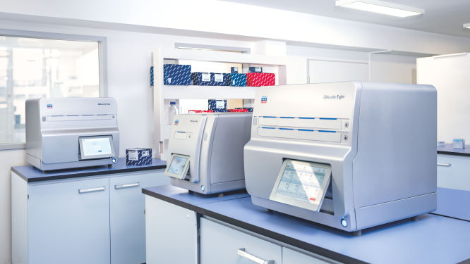
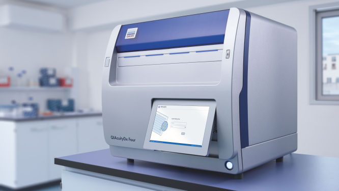
Discover the QIAcuity family of dPCR instruments

Explore the QIAcuity family of dPCR instruments, designed to meet the rigorous demands of biomarker research, translational research and clinical applications. With unmatched precision and operational efficiency, these versatile platforms can transform your scientific and diagnostic efforts.

Transform your research capabilities with QIAcuity digital PCR

QIAcuity is a fully automated digital PCR system that combines precision, efficiency and ease of use. Experience unparalleled accuracy and save resources with high-throughput multiplexing, allowing for the simultaneous detection of up to five genetic targets. A seamless transition from existing qPCR workflows ensures minimal disruption while significantly enhancing data quality and throughput.

Streamline your clinical PCR workflows with QIAcuityDx

QIAcuityDx is tailored for IVD applications. This fully automated system enhances diagnostic precision and operational efficiency by reducing hands-on time and ensuring accurate detection and quantification of important genetic variations. Easily develop your own assay menu* by using QIAcuityDx utility mode and IVD medical device consumables, reagents and software.

*FDA ‘Medical Devices; Laboratory Developed Tests’ final rule, May 6, 2024 and European Union regulation requirements on ‘In-House Assays’ (Regulation (EU) 2017/746 -IVDR- Art. 5(5))

Frequently asked questions

Discover key insights into genes and variants relevant to CNS cancers and neoplasms research and how they can be detected using digital PCR
How do dPCR LNA Mutation Assays benefit cancer researchers?
dPCR LNA Mutation Assays offer significant advantages to cancer researchers working on precise and sensitive mutation detection. These assays are specifically designed for use with the QIAcuity Digital PCR System and are enhanced with Locked Nucleic Acid (LNA) technology. This enhancement greatly improves the specificity and sensitivity of mutation detection, making it possible to identify DNA sequence mutations at very low abundance, with a sensitivity as fine as 0.1% in a single nanoplate well.

The key benefits of dPCR LNA Mutation Assays for cancer researchers include:
  • High precision and sensitivity: The use of duplex, hydrolysis probe-based assays allows for highly precise detection of mutations. The presence of both mutant and wild-type probes in the same reaction ensures that researchers can detect and quantify minor genetic variations with great accuracy, crucial for studies in heterogeneous cancer samples where only a few cells may carry the mutation.
  • Enhanced specificity: The integration of LNA into the probes increases the binding affinity and specificity towards the target sequences, minimizing the risk of non-specific bindings and improving the overall reliability of the assays.
  • Multiplexing capability: Each assay is capable of detecting mutations using two different fluorescent dye combinations, allowing for the simultaneous analysis of mutant and wild-type alleles within the same reaction. This multiplexing ability is particularly useful in applications requiring the analysis of multiple targets, such as assessing co-occurring mutations in cancer.
  • Flexibility in sample analysis: By dividing the reaction across multiple wells, even greater sensitivity can be achieved, facilitating the detection of extremely rare mutations. This is especially valuable in cancer research, where detecting low-frequency mutations can inform prognosis and treatment strategies.
  • Streamlined workflow: Supplied in a single-tube format with ready-to-use primer pairs and probes, these assays simplify the experimental setup, enabling efficient and straightforward integration into existing research workflows.
What role does the TP53 gene play in CNS cancer?
TP53 (Tumor Protein p53) is a crucial tumor suppressor gene that encodes the p53 protein, which plays a vital role in regulating the cell cycle, DNA repair and apoptosis. In brain tumors and particularly glioblastoma (GBM), TP53 mutations are common and contribute significantly to tumor development and progression. The p53 protein acts as a guardian of the genome, responding to DNA damage by either repairing the damage or initiating cell death if the damage is irreparable. It interacts with various proteins involved in cell cycle control, such as MDM2, which regulates p53 levels through a feedback loop. Loss of p53 function due to mutations leads to uncontrolled cell proliferation, genomic instability and resistance to apoptosis, all of which contribute to the aggressive nature of brain malignancies. The TP53 variants below play a critical role by disrupting the normal tumor suppressor functions of p53.
  • TP53 c.743G>A / R248Q: A DNA point mutation from guanine (G) to adenine (A) at nucleotide position 743 (c.743G>A) results in the substitution of arginine (R) with glutamine (Q) at position 248 of the TP53 protein (R248Q). This mutation occurs in the DNA-binding domain of p53, impairing its ability to bind to DNA and activate transcription of target genes involved in cell cycle arrest and apoptosis. The R248Q mutation leads to loss of tumor suppressor function, contributing to uncontrolled cell proliferation and survival, which are hallmarks of cancer. This variant is one of the most common TP53 mutations found in glioblastomas, significantly impacting disease progression and treatment response.
  • TP53 c.818G>A / R273H: A DNA point mutation from guanine (G) to adenine (A) at nucleotide position 818 (c.818G>A) results in the substitution of arginine (R) with histidine (H) at position 273 of the TP53 protein (R273H). This mutation affects the DNA-binding domain of p53, reducing its ability to regulate genes involved in DNA repair, cell cycle control and apoptosis. The R273H mutation leads to a dominant-negative effect, where the mutant p53 protein interferes with the function of any remaining wild-type p53, further promoting tumorigenesis. This variant is frequently observed in CNS cancer and is associated with poor prognosis and resistance to chemotherapy.
  • TP53 c.524G>A / R175H: A DNA point mutation from guanine (G) to adenine (A) at nucleotide position 524 (c.524G>A) results in the substitution of arginine (R) with histidine (H) at position 175 of the TP53 protein (R175H). This mutation occurs in the core DNA-binding domain, disrupting the structural integrity of p53 and its ability to bind DNA. The R175H mutation leads to loss of tumor suppressor activity, allowing cells with DNA damage to proliferate unchecked. This variant is commonly found in brain tumors and contributes to the aggressive nature of the disease and resistance to standard treatments.
  • TP53 c.844C>T / R282W: A DNA point mutation from cytosine (C) to thymine (T) at nucleotide position 844 (c.844C>T) results in the substitution of arginine (R) with tryptophan (W) at position 282 of the TP53 protein (R282W). This mutation affects the DNA-binding domain, impairing p53's ability to regulate target genes involved in cell cycle arrest and apoptosis. The R282W mutation results in a loss of function, contributing to the development and progression of CNS cancer by allowing cells to evade growth control mechanisms. This variant is associated with poor clinical outcomes and resistance to chemotherapy.
  • TP53 c.1010G>A / E337K: A DNA point mutation from guanine (G) to adenine (A) at nucleotide position 1010 (c.1010G>A) results in the substitution of glutamic acid (E) with lysine (K) at position 337 of the TP53 protein (E337K). This mutation occurs in the oligomerization domain, affecting p53's ability to form tetramers, which are necessary for its full tumor suppressor activity. The E337K mutation leads to a partial loss of function, contributing to the development and progression of CNS cancer by impairing p53-mediated cell cycle arrest and apoptosis. This variant is less common but still significant in the context of brain tumor pathogenesis.
What is the function of the EGFR gene in brain tumor progression?
EGFR (Epidermal Growth Factor Receptor) is a proto-oncogene that encodes a receptor tyrosine kinase involved in the regulation of cell growth, survival and differentiation. In brain malignancies, particularly glioblastoma, EGFR mutations and amplifications are frequently observed and are associated with aggressive tumor behavior and poor prognosis. The EGFR protein interacts with various downstream signaling pathways, including the PI3K/AKT and MAPK/ERK pathways, to promote cell proliferation and survival. Overexpression or mutation of EGFR leads to continuous activation of these pathways, driving tumor growth and resistance to apoptosis. Targeting EGFR with specific inhibitors is a therapeutic strategy in EGFR-mutant glioblastomas.
  • EGFR c.2573T>G / L858R: A DNA point mutation from thymine (T) to guanine (G) at nucleotide position 2573 (c.2573T>G) results in the substitution of leucine (L) with arginine (R) at position 858 of the EGFR protein (L858R). This mutation occurs in the tyrosine kinase domain of EGFR, leading to constitutive activation of the receptor. The L858R mutation enhances EGFR signaling through the PI3K/AKT and MAPK/ERK pathways, promoting cell proliferation and survival. This variant is commonly found in glioblastomas and is associated with aggressive tumor behavior and poor prognosis. Targeting L858R with specific EGFR inhibitors can be an effective therapeutic strategy.
  • EGFR c.2235_2249del / Exon 19 Deletion: A deletion of 15 base pairs in exon 19 (c.2235_2249del) results in the loss of five amino acids in the EGFR protein. This exon 19 deletion leads to constitutive activation of the EGFR tyrosine kinase domain, enhancing downstream signaling pathways such as PI3K/AKT and MAPK/ERK. The exon 19 deletion is frequently observed in glioblastomas and contributes to tumor growth and resistance to apoptosis. This variant is a target for EGFR inhibitors, which can inhibit the aberrant signaling and reduce tumor progression.
  • EGFR c.2369C>T / T790M: A DNA point mutation from cytosine (C) to thymine (T) at nucleotide position 2369 (c.2369C>T) results in the substitution of threonine (T) with methionine (M) at position 790 of the EGFR protein (T790M). This mutation occurs in the tyrosine kinase domain and is often associated with resistance to first- and second-generation EGFR inhibitors. The T790M mutation increases the affinity of EGFR for ATP, reducing the effectiveness of ATP-competitive inhibitors. It is commonly found in glioblastomas that have developed resistance to initial EGFR-targeted therapies. Newer generation inhibitors, such as osimertinib, are designed to target this mutation.
  • EGFR c.2361G>A / C797S: A DNA point mutation from guanine (G) to adenine (A) at nucleotide position 2361 (c.2361G>A) results in the substitution of cysteine (C) with serine (S) at position 797 of the EGFR protein (C797S). This mutation occurs in the tyrosine kinase domain and is associated with resistance to third-generation EGFR inhibitors, such as osimertinib. The C797S mutation prevents the covalent binding of these inhibitors to the EGFR protein, allowing continued signaling through the PI3K/AKT and MAPK/ERK pathways. This variant is significant in the context of acquired resistance in glioblastomas.
  • EGFR c.2570T>C / L861Q: A DNA point mutation from thymine (T) to cytosine (C) at nucleotide position 2570 (c.2570T>C) results in the substitution of leucine (L) with glutamine (Q) at position 861 of the EGFR protein (L861Q). This mutation occurs in the tyrosine kinase domain and leads to constitutive activation of EGFR, promoting cell proliferation and survival through downstream signaling pathways. The L861Q mutation is less common but still relevant in glioblastomas, contributing to tumor growth and progression. Targeting this variant with specific EGFR inhibitors can be a therapeutic approach.
How does the IDH1 gene influence the development of CNS cancer?
IDH1 (Isocitrate Dehydrogenase 1) encodes an enzyme that is involved in the citric acid cycle, playing a role in cellular metabolism. In brain tumors, particularly lower-grade gliomas and secondary glioblastomas, mutations in IDH1 are common and lead to the production of an oncometabolite called 2-hydroxyglutarate (2-HG). This metabolite interferes with cellular differentiation and promotes tumorigenesis by altering the epigenetic landscape of the cell. IDH1 mutations are associated with distinct metabolic and epigenetic changes that drive tumorigenesis. These mutations are also linked to better prognosis and response to certain therapies compared to IDH1 wild-type gliomas. Some important variants in IGH1 associated with CNS cancer include:
  • IDH1 c.395G>A / R132H: A DNA point mutation from guanine (G) to adenine (A) at nucleotide position 395 (c.395G>A) results in the substitution of arginine (R) with histidine (H) at position 132 of the IDH1 protein (R132H). This mutation occurs at the active site of the enzyme, leading to the production of the oncometabolite 2-hydroxyglutarate (2-HG). The accumulation of 2-HG interferes with cellular differentiation and promotes tumorigenesis by altering the epigenetic landscape. The R132H mutation is the most common IDH1 mutation in gliomas and is associated with better prognosis and response to certain therapies compared to IDH1 wild-type gliomas.
  • IDH1 c.394C>T / R132C: A DNA point mutation from cytosine (C) to thymine (T) at nucleotide position 394 (c.394C>T) results in the substitution of arginine (R) with cysteine (C) at position 132 of the IDH1 protein (R132C). Similar to R132H, this mutation leads to the production of 2-hydroxyglutarate (2-HG), promoting tumorigenesis through epigenetic alterations. The R132C mutation is less common than R132H but still significant in the context of glioma development and progression. It is associated with distinct metabolic and epigenetic changes that drive tumorigenesis.
What impact does the PTEN gene have on CNS cancer?
PTEN (Phosphatase and Tensin Homolog) is a tumor suppressor gene that encodes a phosphatase involved in the negative regulation of the PI3K/AKT signaling pathway. In brain tumors, particularly glioblastoma, PTEN mutations lead to unregulated activation of the PI3K/AKT pathway, promoting cell growth and survival. The PTEN protein dephosphorylates phosphatidylinositol-3,4,5-trisphosphate (PIP3), antagonizing PI3K signaling. Loss of PTEN function results in increased PIP3 levels, continuous activation of AKT and enhanced cell proliferation and survival. PTEN mutations contribute to the aggressive nature of CNS neoplasms and are associated with poor prognosis.
What is the significance of the BRAF gene in CNS cancer?
BRAF (B-Raf Proto-Oncogene, Serine/Threonine Kinase) is a proto-oncogene that encodes a protein kinase involved in the MAPK/ERK signaling pathway, which regulates cell growth, differentiation and survival. In brain tumors, particularly in certain pediatric tumors such as pilocytic astrocytomas and pleomorphic xanthoastrocytomas, BRAF mutations, especially V600E, are common. These mutations lead to constitutive activation of the BRAF kinase, driving continuous MAPK/ERK pathway signaling and promoting tumor growth. BRAF mutations are associated with distinct biological behaviors and can influence the response to targeted therapies, such as BRAF inhibitors.
How do mutations in the PIK3CA gene contribute to CNS cancer?
PIK3CA (Phosphatidylinositol-4,5-Bisphosphate 3-Kinase Catalytic Subunit Alpha) encodes the p110α catalytic subunit of the PI3K enzyme, which is involved in the PI3K/AKT/mTOR signaling pathway. This pathway regulates cell growth, proliferation and survival. In Cbrain tumors, PIK3CA mutations lead to the activation of the PI3K/AKT pathway, promoting tumor growth and survival. The PIK3CA protein interacts with the regulatory subunit p85 and other signaling molecules to mediate its effects. Dysregulation of this pathway due to PIK3CA mutations contributes to cancer progression and resistance to certain therapies. Targeting the PI3K/AKT/mTOR pathway is a potential therapeutic strategy in PIK3CA-mutant CNS cancers.
How do H3F3A gene mutations affect CNS cancer development?
H3F3A (H3 Histone Family Member 3A) encodes a variant of the histone H3 protein, which is involved in the packaging of DNA into chromatin and regulation of gene expression. In brain tumors, particularly in pediatric high-grade gliomas and diffuse midline gliomas, mutations in H3F3A, such as K27M and G34R/V, are characteristic. These mutations alter the chromatin structure and epigenetic landscape, leading to dysregulation of gene expression and promoting tumorigenesis. H3F3A mutations are associated with aggressive tumor behavior and poor prognosis, and they define distinct molecular subgroups of CNS tumors.
How does the ACVR1 gene influence CNS cancer growth?
ACVR1 (Activin A Receptor Type 1) encodes a receptor serine/threonine kinase that is involved in the bone morphogenetic protein (BMP) signaling pathway, which regulates cell growth, differentiation and apoptosis. In brain tumors, particularly in diffuse intrinsic pontine gliomas (DIPG), ACVR1 mutations are common and lead to aberrant BMP signaling. These mutations result in constitutive activation of the ACVR1 receptor, promoting tumor growth and resistance to apoptosis. ACVR1 mutations are associated with distinct biological behaviors and can influence the response to targeted therapies in DIPG.
What role does the NF2 gene play in CNS cancer?
NF2 (Neurofibromin 2) is a tumor suppressor gene that encodes the protein merlin, which is involved in regulating cell growth, adhesion and cytoskeletal organization. In CNS cancer, particularly in meningiomas and schwannomas, NF2 mutations lead to the loss of merlin function, resulting in dysregulated cell growth and adhesion. The NF2 protein interacts with various signaling pathways, including the Hippo pathway, to control cell proliferation and survival. Loss of NF2 function contributes to the development and progression of these tumors, and NF2 mutations are a hallmark of neurofibromatosis type 2, a genetic disorder that predisposes individuals to multiple CNS neoplasms.
What is the involvement of the PTCH2 gene in CNS cancer?
PTCH2 (Patched 2) is a tumor suppressor gene that encodes a receptor involved in the Hedgehog signaling pathway, which regulates cell growth, differentiation and tissue patterning. In brain tumors, particularly in medulloblastomas, PTCH2 mutations lead to dysregulation of the Hedgehog pathway, promoting tumor growth and survival. The PTCH2 protein interacts with the Smoothened (SMO) receptor to inhibit Hedgehog signaling. Loss of PTCH2 function results in continuous activation of the pathway, contributing to the development and progression of medulloblastomas. PTCH2 mutations are associated with distinct molecular subgroups of medulloblastomas and can influence the response to targeted therapies.

Disclaimers

dPCR LNA Mutation Assays are intended for molecular biology applications. These products are not intended for the diagnosis, prevention, or treatment of a disease.

The QIAcuity is intended for molecular biology applications. This product is not intended for the diagnosis, prevention or treatment of a disease. Therefore, the performance characteristics of the product for clinical use (i.e., diagnostic, prognostic, therapeutic or blood banking) is unknown.

The QIAcuityDx dPCR System is intended for in vitro diagnostic use, using automated multiplex quantification dPCR technology, for the purpose of providing diagnostic information concerning pathological states.

QIAcuity and QIAcuityDx dPCR instruments are sold under license from Bio-Rad Laboratories, Inc. and exclude rights for use with pediatric applications. The QIAcuityDx medical device is currently under development and will be available in 20 countries in H2 2024.