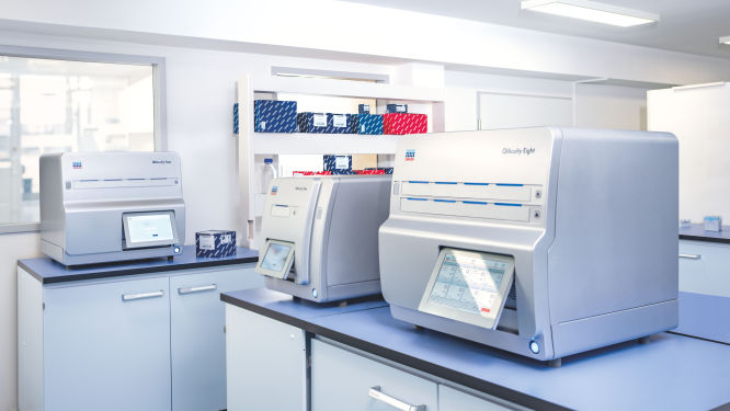
hero image

Digital PCR assays for colorectal cancer gene variants

Order ready-to-use dPCR assays

Revolutionizing colorectal cancer research with precision dPCR assays

The biological complexity and frequent late diagnoses of colorectal cancer pose significant challenges to researchers. Each case of colorectal cancer can vary significantly in terms of genetic mutations and clinical presentation, which necessitates precise characterization of gene variants to tailor effective treatments.

Digital PCR (dPCR) emerges as a vital tool in this context because it offers highly sensitive and accurate quantification of genetic material. This precision makes it possible to detect even low levels of genetic mutations that could be crucial for understanding the specific pathways involved in individual cases, thereby facilitating research into more targeted and effective therapies.

Explore colorectal cancer related dPCR assays by gene

A variety of gene variants are associated with the development and progression of colorectal cancer. Key mutations in genes such as APC, KRAS, TP53 and BRAF play significant roles in driving tumor growth and influencing resistance to therapies. Specifically, APC mutations are common in the early stages of carcinogenesis, marking it as an essential target for early detection. Meanwhile, KRAS mutations critically affect the tumor's responsiveness to certain chemotherapeutic agents and targeted therapies, underscoring the importance of personalized treatment plans.

Our collection of dPCR LNA Mutation Assays provides researchers with essential tools for in-depth analysis of these and other pivotal genetic changes. These assays facilitate precise quantification and characterization, contributing to the advancement of targeted research and therapeutic development.

The table below includes COSMIC Variant IDs (COSV), as they provide a unique identifier for each distinct gene mutation and ensure precise reference to your variant of interest.

Discover the QIAcuity family of dPCR instruments

Explore the QIAcuity family of dPCR instruments, designed to meet the rigorous demands of biomarker research, translational research and clinical applications. With unmatched precision and operational efficiency, these versatile platforms can transform your scientific and diagnostic efforts.
Transform your research capabilities with QIAcuity digital PCR

Transform your research capabilities with QIAcuity digital PCR

QIAcuity is a fully automated digital PCR system that combines precision, efficiency and ease of use. Experience unparalleled accuracy and save resources with high-throughput multiplexing, allowing for the simultaneous detection of up to five genetic targets. A seamless transition from existing qPCR workflows ensures minimal disruption while significantly enhancing data quality and throughput.
Explore QIAcuity
Streamline your clinical PCR workflows with QIAcuityDx

Streamline your clinical PCR workflows with QIAcuityDx

QIAcuityDx is tailored for IVD applications. This fully automated system enhances diagnostic precision and operational efficiency by reducing hands-on time and ensuring accurate detection and quantification of important genetic variations. Easily develop your own assay menu* by using QIAcuityDx utility mode and IVD medical device consumables, reagents and software.
Explore QIAcuityDx

*FDA ‘Medical Devices; Laboratory Developed Tests’ final rule, May 6, 2024 and European Union regulation requirements on ‘In-House Assays’ (Regulation (EU) 2017/746 -IVDR- Art. 5(5))

Frequently asked questions

Discover key insights into genes and variants critical for colorectal cancer research and how they can be detected using digital PCR

How do dPCR LNA Mutation Assays benefit cancer researchers?

dPCR LNA Mutation Assays offer significant advantages to cancer researchers working on precise and sensitive mutation detection. These assays are specifically designed for use with the QIAcuity Digital PCR System and are enhanced with Locked Nucleic Acid (LNA) technology. This enhancement greatly improves the specificity and sensitivity of mutation detection, making it possible to identify DNA sequence mutations at very low abundance, with a sensitivity as fine as 0.1% in a single nanoplate well.

The key benefits of dPCR LNA Mutation Assays for cancer researchers include:
  • High precision and sensitivity: The use of duplex, hydrolysis probe-based assays allows for highly precise detection of mutations. The presence of both mutant and wild-type probes in the same reaction ensures that researchers can detect and quantify minor genetic variations with great accuracy, crucial for studies in heterogeneous cancer samples where only a few cells may carry the mutation.
  • Enhanced specificity: The integration of LNA into the probes increases the binding affinity and specificity towards the target sequences, minimizing the risk of non-specific bindings and improving the overall reliability of the assays.
  • Multiplexing capability: Each assay is capable of detecting mutations using two different fluorescent dye combinations, allowing for the simultaneous analysis of mutant and wild-type alleles within the same reaction. This multiplexing ability is particularly useful in applications requiring the analysis of multiple targets, such as assessing co-occurring mutations in cancer.
  • Flexibility in sample analysis: By dividing the reaction across multiple wells, even greater sensitivity can be achieved, facilitating the detection of extremely rare mutations. This is especially valuable in cancer research, where detecting low-frequency mutations can inform prognosis and treatment strategies.
  • Streamlined workflow: Supplied in a single-tube format with ready-to-use primer pairs and probes, these assays simplify the experimental setup, enabling efficient and straightforward integration into existing research workflows.

How is KRAS involved in colorectal cancer?

KRAS (Kirsten rat sarcoma viral oncogene homolog) is a gene encoding a GTPase that functions as a molecular switch within the RAS/MAPK signaling pathway. This pathway regulates crucial cellular processes including cell division, differentiation and apoptosis. In colorectal cancer, mutations in the KRAS gene are common and constitute significant oncogenic drivers that lead to the constitutive activation of downstream signaling pathways such as MAPK and PI3K, promoting cellular proliferation and survival independent of external growth signals. The most frequent mutations in KRAS involve codons 12 and 13, which result in a protein that is constitutively active, unable to hydrolyze GTP and perpetually engaged in signaling. This aberrant activity bypasses the normal regulatory mechanisms that would otherwise control cell growth and apoptotic processes, facilitating the development and progression of malignant tumors.

KRAS mutations are associated with resistance to EGFR-targeted therapies such as cetuximab and panitumumab, which are used in the treatment of metastatic colorectal cancer. This resistance arises because the mutated KRAS gene continues to activate the downstream signaling irrespective of EGFR inhibition, rendering these therapies ineffective. The presence of KRAS mutations in a tumor is considered a hallmark of poor prognosis and is integral to therapeutic decision-making in clinical practice, guiding the selection of treatment regimens that do not rely on EGFR blockade.

  • KRAS c.35G>A / G12D: This involves a point mutation from glycine (G) to aspartate (D) at codon 12 of the KRAS gene. The G12D mutation leads to the production of a constitutively active KRAS protein that no longer requires growth factor signals to promote cell division, contributing to uncontrolled cell growth and tumor development. This mutation is common in colorectal cancer and is associated with resistance to anti-EGFR therapies, making it a crucial marker for treatment planning.
  • KRAS c.35G>T / G12V: A point mutation where valine (V) replaces glycine (G) at codon 12. Similar to G12D, the G12V mutation results in an oncogenic KRAS protein that drives continuous cell proliferation. It is particularly detrimental as it is associated with poor prognosis and survival in colorectal cancer patients, and it also influences the efficacy of targeted therapeutic interventions.

What role does APC play in colorectal cancer?

APC (Adenomatous Polyposis Coli) encodes a tumor suppressor protein playing a pivotal role in the regulation of the WNT signaling pathway, which is essential for cellular proliferation, differentiation and apoptosis. In colorectal cancer, mutations in the APC gene are among the most common early events, leading to the constitutive activation of the WNT pathway. This results in the accumulation of β-catenin in the nucleus and the activation of WNT target genes that promote uncontrolled cell growth and the eventual development of adenomatous polyps. The canonical function of the APC protein includes the formation of a destruction complex involving several other proteins like GSK-3β, AXIN and CK1, which facilitate the phosphorylation and subsequent proteasomal degradation of β-catenin, thereby preventing its oncogenic activity. Loss of APC function disrupts this complex, leading to an aberrant accumulation of β-catenin and enhancement of oncogenic transcription. Some specific APC variants include:
  • APC c.3920A>G / I1307K: The I1307K variant of the APC gene is a missense mutation involving the substitution of lysine (K) for isoleucine (I) at amino acid position 1307. This mutation increases the likelihood of erroneous DNA replication due to the altered protein structure, particularly in the β-catenin binding and degradation domain. While the I1307K mutation does not create an immediate cancerous condition, it significantly enhances the propensity for developing adenomatous polyps, which are precursors to colorectal cancer. It is especially prevalent among Ashkenazi Jews, conferring a moderate increase risk of developing the disease.
  • APC c.3949G>C / E1317Q: Another missense mutation in the APC gene, where glutamine (Q) replaces glutamic acid (E) at position 1317. Similar to I1307K, this mutation subtly affects the protein's ability to bind and regulate β-catenin, though its direct link to colorectal cancer risk is less pronounced than other APC mutations. It predisposes carriers to multiple colorectal adenomas, much like I1307K.

How does MLH1 influence colorectal cancer?

MLH1 (MutL Homolog 1) encodes a key protein in the DNA mismatch repair (MMR) system, crucial for correcting DNA replication errors and maintaining genomic stability. In colorectal cancer, loss of MLH1 function through mutations or epigenetic silencing leads to microsatellite instability (MSI), which accelerates mutation accumulation and cancer progression. This is particularly significant in Lynch syndrome, a condition that predisposes individuals to colorectal and other cancers. MSI-high colorectal cancers, often resulting from MLH1 deficiency, tend to respond well to immunotherapies due to their high mutational burden, making them more visible to the immune system. Moreover, the status of MLH1 is critical for diagnosing Lynch syndrome, guiding treatment choices and has implications for prognosis and therapeutic resistance.
  • MLH1 c.655A>G / p.Lys219Glu: This mutation involves a nucleotide change from adenine (A) to guanine (G) at position 655, resulting in the substitution of glutamic acid (E) for lysine (K) at amino acid position 219 in exon 8. The p.Lys219Glu mutation alters the protein's structure necessary for its interaction within the mismatch repair complex, particularly affecting its binding efficiency with PMS2. This disruption leads to compromised DNA repair functionality, increasing the rate of mutation accumulation across the genome. The resultant microsatellite instability is a hallmark of Lynch syndrome-associated colorectal cancer, influencing both tumor behavior and response to treatment.
  • MLH1 c.790+1G>A: This splice site mutation involves a change from guanine (G) to adenine (A) at the first nucleotide of intron 9 (790+1). This alteration affects the canonical splice site, leading to aberrant splicing of the MLH1 mRNA. The incorrect splicing results in a malformed protein that lacks critical regions necessary for its function in the mismatch repair process. The defective protein cannot adequately interact with other MMR proteins, resulting in microsatellite instability (MSI). This instability is characteristic of Lynch syndrome, which predisposes individuals to early-onset colorectal cancer and influences therapeutic responses, particularly enhancing effectiveness of immunotherapies.
  • MLH1 c.677+3A>T: Another splice site mutation, this change occurs in the third nucleotide of intron 7 from adenine (A) to thymine (T). This mutation disrupts normal splicing mechanisms, typically resulting in the exclusion of exon 8 from the mRNA. The resulting truncated protein lacks essential domains required for its function, impairing the MMR system and leading to increased mutational burden within the cell. This variant is frequently associated with Lynch syndrome, predisposing carriers to multiple cancers including colorectal cancer, and has significant implications for prognosis and treatment strategy.
  • MLH1 p.G67R: This missense mutation involves the substitution of arginine (R) for glycine (G) at codon 67, located in exon 3 of the MLH1 gene. The p.G67R mutation affects a region critical for the structural integrity and proper function of the MLH1 protein. By altering the protein’s ability to form a stable complex with PMS2, this mutation compromises the effectiveness of the mismatch repair system. This deficiency leads to MSI-high colorectal cancers, typically associated with better outcomes when treated with immune checkpoint inhibitors but may confer resistance to certain chemotherapeutic agents.

What is the significance of MSH2 in colorectal cancer development?

MSH2 (MutS Homolog 2) is integral to the DNA mismatch repair (MMR) system, working with MLH1 to correct replication errors. Mutations in MSH2 lead to MMR deficiency and the associated microsatellite instability (MSI) in colorectal cancer, significantly fostering cancer development. This deficiency is notably researched in the context of cancer biology and genetics. Studying MSH2 mutations offers insights into the molecular pathways that predispose to colorectal and other related cancers. Research on MSH2 can also help in developing novel therapeutic approaches, such as enhancing the effectiveness of immunotherapies in tumors with MMR deficiency.

How is MSH6 implicated in colorectal cancer mechanisms?

MSH6 (MutS Homolog 6) works alongside MLH1 and MSH2 in the DNA mismatch repair system, ensuring genomic stability. Mutations in MSH6 disrupt this repair process, leading to microsatellite instability and MMR deficiency, key factors in the development of colorectal cancer. Research into MSH6 mutations provides valuable data on the molecular mechanisms of cancer progression and can inform the development of strategies to target these pathways. Additionally, understanding the role of MSH6 in genetic instability can help in the design of targeted therapies, particularly immunotherapies, which have shown increased efficacy in MSI-high cancers.

How is PIK3CA involved in colorectal cancer?

PIK3CA encodes the p110α catalytic subunit of phosphatidylinositol 3-kinase (PI3K), a lipid kinase that is a critical component of the PI3K/AKT/mTOR pathway, a signaling axis that plays a central role in cellular processes such as growth, proliferation and survival. In colorectal cancer, mutations in PIK3CA are frequently observed and contribute to the pathogenesis of the disease through the enhancement of this signaling pathway. The involvement of PIK3CA mutations in colorectal cancer is also associated with specific clinical outcomes and can influence response to certain therapies. For instance, these mutations have been linked to resistance to anti-EGFR therapies in KRAS wild-type cancers, suggesting a complex interplay between PI3K and other pathways in colorectal cancer. Targeting the PI3K pathway, therefore, represents a promising therapeutic strategy and has led to the development and approval of specific inhibitors that can potentially improve treatment outcomes for patients harboring PIK3CA mutations.
  • PIK3CA c.1633G>A / E545K: This mutation is characterized by a nucleotide change from guanine (G) to adenine (A) at position 1633, resulting in the substitution of lysine (K) for glutamic acid (E) at amino acid position 545 in exon 9. E545K affects the helical domain of the PI3K catalytic subunit, leading to activation of the PI3K/AKT signaling pathway independent of upstream signals. The increased kinase activity from this mutation results in constitutive pathway activation, stimulating cellular growth and proliferation while inhibiting apoptosis. This autonomous signaling supports the survival and expansion of malignant cells, making E545K a common mutation in colorectal cancer that frequently co-occurs with mutations like KRAS and complicates the efficacy of chemotherapeutic agents.
  • PIK3CA c.3140A>G / H1047R: The H1047R mutation, involving a nucleotide substitution from adenine (A) to guanine (G) at position 3140 in the PIK3CA gene, results in the replacement of histidine (H) with arginine (R) at amino acid position 1047 in exon 20. H1047R occurs in the kinase domain of PIK3CA, resulting in increased lipid kinase activity that leads to enhanced downstream AKT signaling. This constitutive activation promotes oncogenesis by fostering cell growth and survival while also playing a role in therapeutic resistance. H1047R is a potent oncogenic driver in colorectal cancer, often associated with poor response to targeted therapies, emphasizing its clinical significance in treatment strategy considerations.

How does PMS2 involvement impact colorectal cancer?

PMS2 (Postmeiotic Segregation Increased 2) collaborates with MLH1 to correct DNA replication errors within the mismatch repair system. Mutations in PMS2 lead to MMR deficiency and microsatellite instability, contributing to colorectal cancer development. Research on PMS2 mutations is crucial for understanding their role in cancer biology and in informing therapeutic development. Studies on PMS2 can aid in elucidating the complex interactions within the MMR pathway and their effects on tumor behavior, potentially guiding therapeutic innovation, especially for treatments that leverage the immune response in MSI-high tumors.

What is the role of SMAD4 in the progression of colorectal cancer?

SMAD4 (SMAD Family Member 4) regulates the TGF-beta signaling pathway, crucial for cell growth, differentiation and apoptosis. Mutations in SMAD4 disrupt this pathway, contributing to colorectal cancer progression. Research on SMAD4 mutations is vital for understanding the molecular dynamics of tumor progression and the interaction between TGF-beta signaling and other critical pathways like WNT and PI3K/AKT. This knowledge is instrumental in developing novel therapeutic approaches targeting these pathways, which could improve treatment outcomes for patients with altered SMAD4 function.

In what way is TP53 involved in colorectal cancer?

TP53 (Tumor Protein p53) is pivotal in regulating the cell cycle, DNA repair and apoptosis. Mutations in TP53 impair its function, allowing colorectal cancer cells to proliferate despite DNA damage, thus promoting tumor progression. These mutations frequently result in a loss of protein function, contributing directly to tumor development by failing to halt cell division in response to genetic damage. Understanding TP53 mutations is crucial for exploring the mechanisms of tumor aggression and therapy resistance. This knowledge can guide the development of targeted therapies aiming to compensate for the loss of p53 function or enhance the efficacy of existing treatments.

Disclaimers

dPCR LNA Mutation Assays are intended for molecular biology applications. These products are not intended for the diagnosis, prevention, or treatment of a disease.

The QIAcuity is intended for molecular biology applications. This product is not intended for the diagnosis, prevention or treatment of a disease. Therefore, the performance characteristics of the product for clinical use (i.e., diagnostic, prognostic, therapeutic or blood banking) is unknown.

The QIAcuityDx dPCR System is intended for in vitro diagnostic use, using automated multiplex quantification dPCR technology, for the purpose of providing diagnostic information concerning pathological states.

QIAcuity and QIAcuityDx dPCR instruments are sold under license from Bio-Rad Laboratories, Inc. and exclude rights for use with pediatric applications. The QIAcuityDx medical device is currently under development and will be available in 20 countries in H2 2024.